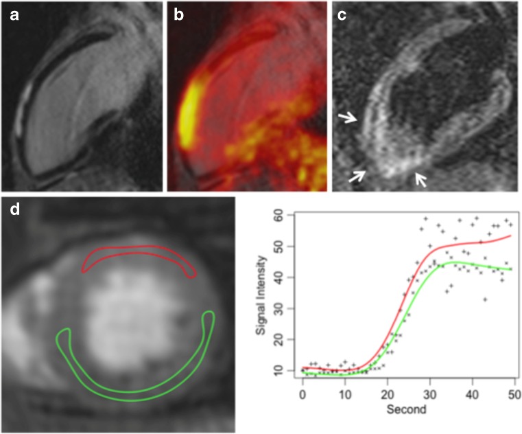
Fig. 5.
18F-FDG PET/MRI in a patient with acute viral myocarditis caused by parvovirus B19. (A) Late-gadolinium-enhanced MRI long-axis view demonstrating typical subepicardial enhancement in the anterior left ventricular wall that was in excellent spatial agreement with increased 18F-FDG uptake on fused images (B). (C) T2-weighted images revealed an oedema in the LV anterior wall. (D) Dynamic perfusion imaging revealed hyperaemia in the LV anterior wall. (With kind permission from Ref 50-Nensa, Poeppel 2014)

