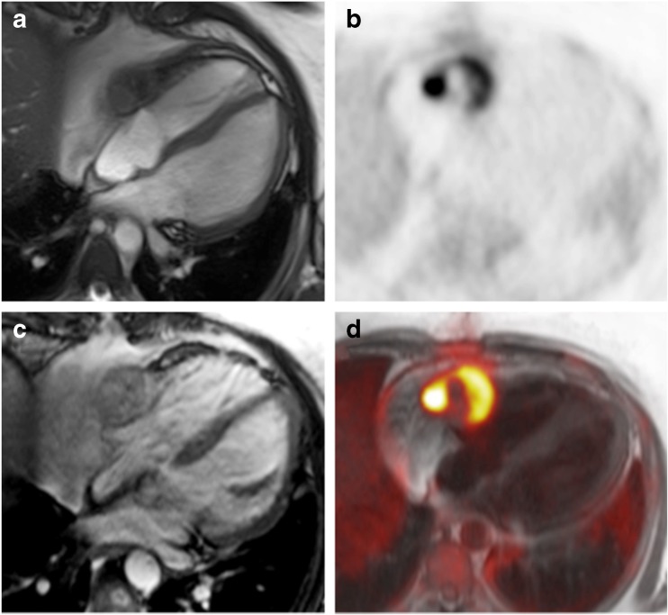
Fig. 6.
(A) Cine MR image of an angiosarcoma infiltrating the free wall of the right ventricle and atrium with adjacent pericardial effusion. (B) 18F-FDG PET shows intense but heterogeneous 18F-FDG uptake within the tumour and otherwise suppressed myocardial 18F-FDG uptake by the use of a high-fat low-carbohydrate protein-permitted diet. (C) The tumour demonstrates heterogeneous and overall moderate enhancement on T1-weighted MRI after intravenous application of gadolinium-based contrast agent. (D) Fused images show excellent spatial agreement between PET and MRI

