
Fig. 7.
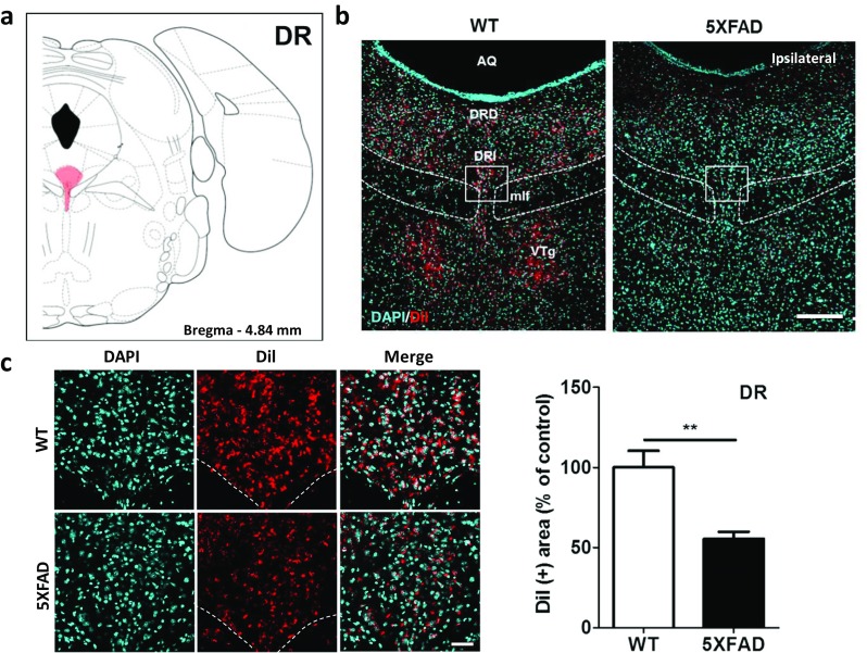
The innervation of the hippocampus from the DR was significantly decreased in the 5XFAD mice compared to the wild-type mice. a Diagram of a mouse brain atlas illustrating the location of the DR at bregma − 4.84 mm. b Representative figures of DiI-positive somata in the midbrain raphe nuclei. DiI-positive cells were mainly observed in the DR and VTg. Scale bar = 200 μm. c Quantification of the DiI-positive area in the DRI. Scale bar = 50 μm. **p < 0.01 indicates significant differences between the groups. AQ, cerebral aqueduct; DR, dorsal raphe; DRD, dorsal raphe nucleus, dorsal part; DRI: dorsal raphe nucleus, interfascicular part; mlf, medial longitudinal fasciculus; VTg, ventral tegmental nucleus