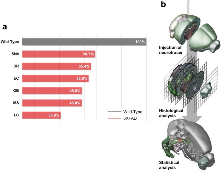
Fig. 9.
Schematic drawing of the decreased inputs into the hippocampus of 5XFAD mice. a A quantitative analysis confirmed that the number of DiI-positive cells was significantly decreased in the EC, MS, LC, DR, SNc, and OB of 11.5-month-old 5XFAD mice compared with wild-type littermate mice. b The experimental design for examining the decreased hippocampal connectivity in an animal model of AD. DiI was injected into the hippocampus of the 5XFAD mice. The tracer was taken up by axonal terminals within the hippocampus and then transported to remote regions through the axons of the neurons. Four days after the injections, histochemical analyses were conducted to show the DiI-positive hippocampal afferents

