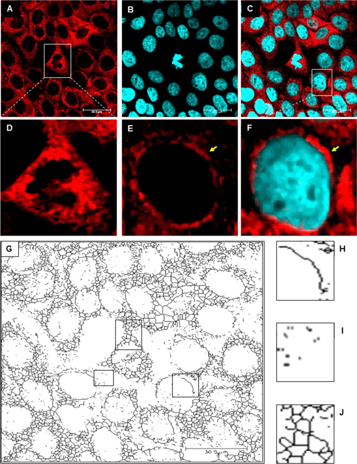
Fig. 2.
The mitochondrial network in uninfected control HaCaT cells. A-F. Immunofluorescence staining of mitochondria (red fluorescence) and the nucleus (blue fluorescence). A yellow arrow indicates tubular mitochondria. G. The original image was processed using “unsharp mask”, “CLAHE”, “median”, “binarize” and “skeletonize” for detection different mitochondrial shapes and structures. H. Tubular mitochondria. I. Punctate mitochondria. J. Branched mitochondrial network

