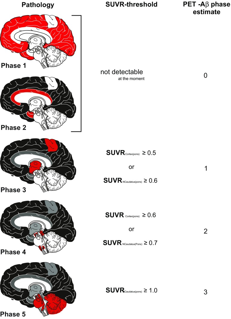
Fig. 2.
SUVR-based protocol for determination of PET-Aβ phase estimates and its link to the pathologically determined phases of Aβ-plaque deposition [36]. Although Aβ-phases 1 and 2 cannot be detected by [18F]flutemetamol PET, cases in Aβ-phase 3 can be identified within one group, i.e. PET-Aβ phase estimate 1, cases in Aβ-phases 4 and 5, respectively, in two further groups, i.e. PET-Aβ phase estimates 2 and 3. The red mark in the schematic representation of the Aβ-phases covers the area in which newly developed plaques in a given phase will develop. This does not mean that the entire red marked field is filled up with Aβ-plaques but that the first small groups of plaques in a given phase of Aβ-depositions can be found there. SUVRCortex(pons) = SUVRcort; SUVRNCaudatus(pons) = SUVRcaud. Picture elements of this figure were taken from a previously published figure [35] and reused with permission

