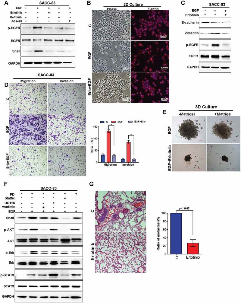
Figure 5.

Targeting EGFR inhibits EGF-induced migration and invasion capabilities of SACC both in vitro and in vivo.
(A) SACC-83 cells were pre-treated with either erlotinib, geftinib or AG1478 for 1 h before stimulation with EGF for 2 h. The expression of Snail and the activation of EGFR were examined by western blotting.(B) The morphology change of SACC-83 cells treated with or without EGF and erlotinib visualized by phase-contrast microscopy, or the cells were stained with the F-actin.(C) Western blot analysis of E-cadherin, vimentin, p-EGFR and EGFR levels from SACC-83 cells treated with or without EGF and erlotinib for 72 hours.(D) Representative images of migration and invasion by SACC-83 cells treated with or without EGF and erlotinib for 72 hours. And graphic representation of percent migrated and invasion cells from 3 separate experiments with mean ± SD percent indicated. * indicates a p < 0.05.(E) 3D morphology of SACC-83 cells treated with or without EGF and erlotinib. Scale bar = 100 μm.(F) Western blot analysis of Snail, p-AKT, AKT, -ERK, ERK, p-STAT3, STAT3 from SACC-83 cells pretreated with various inhibitors for 1 hr followed by stimulation with EGF for 2 hr.(G) SACC-LM cells treated with or without erlotinib for 72 hours were injected into the tail vein of nude mice. Histopathologic analysis shows small metastatic nodules in lung tissues. Graphic representation of the area of lung metastases with mean ± SD; n = 5.
