
Figure 2.
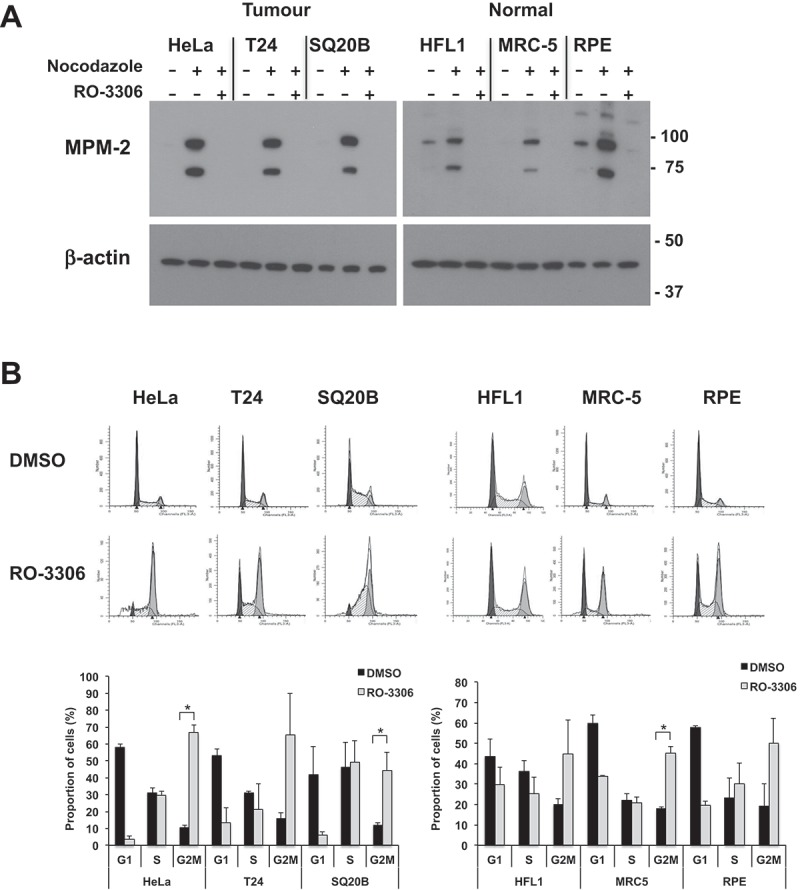
RO-3306 treatment causes inhibition of downstream signaling and cell cycle arrest in both tumor and normal cells. (a) Western blot stained with the MPM-2 antibody, recognizing phosphorylated CDK1 substrates. Cultures were enriched for mitotic cells by overnight nocodazole treatment followed by 2 h treatment with 5 µM RO-3306 in the continuous presence of nocodazole. Both floating and adherent cells were lysed and subjected to immunoblotting. Image is representative of three independent experiments. For clarity, a longer exposure is shown for the normal cell lysates; a comparison of different exposure times is shown in Supplementary Figure 2. (b) Cell cycle profile of tumor and normal cells treated with RO-3306. Cells were seeded in 6-well plates and treated with 5 µM RO-3306 for 20 h. Cells were lifted, fixed in 70% ethanol and stained with propidium iodide for analysis by flow cytometry. Representative histograms are shown (dark grey: G1, white: S; light grey: G2/M). Graph shows percentages in each cell cycle phase determined through curve fitting with ModFit and show the mean +/- SD from two independent experiments. Significant increases in G2/M proportion after RO-3306 treatment are indicated (* p < 0.05).