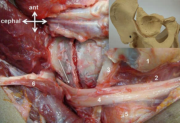
Fig. 19.

Showing kinking of the sciatic nerve over the edge of the stable ilium (arrow) with substantial medial shifting of the acetabular fragment. 1 = greater trochanter, 2 = quadratus femoris, 3 = obturator externus, 4 = sciatic nerve, 5 = ischium, and 6 = reflected piriformis.
