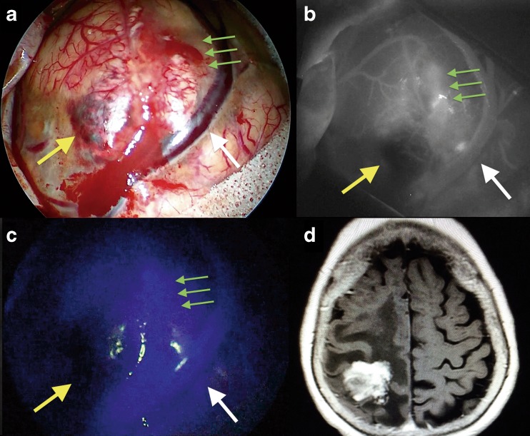
Fig. 9.
The intraoperative findings during a parietal lobe metastasis of ovarian carcinoma removal via a right parietal approach. a Microscopic view before tumor removal under white light; yellow arrow shows the necrotic part of the lesion while the green arrows show the blood-brain barrier disruption. b The same surgical field as in a under indocyanine microscopic integrated view; yellow arrow shows the necrotic part of the lesion while the green arrows show the blood-brain barrier disruption. c The same surgical field as in b under endoscopic assisted view and near-infrared light after injection of ICG; yellow arrow shows the necrotic part of the lesion while the green arrows show the blood-brain barrier disruption

