Fig. 6.
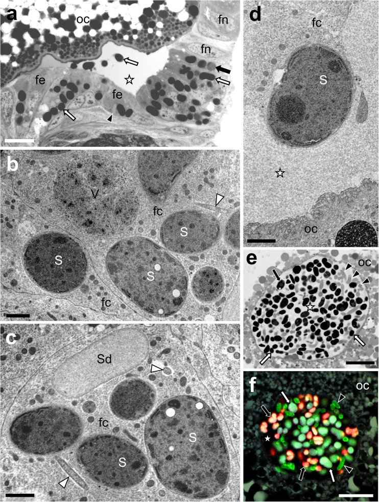
Consecutive stages of transovarial transmission of symbiotic bacteria in Ommatidiotus dissimilis. a The migration of the symbiotic bacteria through the follicular epithelium surrounding the posterior pole of the terminal oocyte to the previtelline space. Bacterium Sulcia (white arrow); bacterium Vidania (black arrow); Sodalis-like bacterium (black arrowhead); follicular epithelium (fe); nucleus of the follicular cell (fn); oocyte (oc); perivitelline space (white asterisk). LM, scale bar = 20 μm. b, c Symbiotic bacteria in the cytoplasm of the follicular cells (fc). Bacterium Sulcia (S); bacterium Vidania (V); Sodalis-like bacterium (Sd); small, rod-shaped bacterium (white arrowhead). TEM, scale bar = 2 μm. d Bacterium Sulcia (S) migrating from the follicular cell to the perivitelline space. Follicular cell (fc); oocyte (oc); perivitelline space (white asterisk). TEM. Scale bar = 2 μm. e A “symbiont ball” in the deep depression of the oolemma. Bacterium Sulcia (white arrow); bacterium Vidania (black arrow); Sodalis-like bacterium (black arrowhead); oocyte (oc); perivitelline space (white asterisk). LM, scale bar = 20 μm. f FISH detection of bacteria Sulcia (white arrow), Vidania (black arrow), and Sodalis (black arrowhead) in the “symbiont ball.” Oocyte (oc); perivitelline space (white asterisk). Confocal microscope, scale bar = 20 μm

