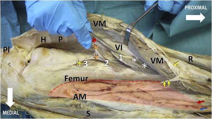
Fig. 1.
Photograph showing the medial view of the distal two-thirds of a right thigh. The vastus medialis (VM), together with its insertion into the vastus intermedius (VI), is lifted from its hammock-like origin (red transparent shading), the medial lip of the linea aspera, and the medial supracondylar line (red dotted line), thereby revealing the knee joint cavity, the articularis genus (indicated by the white numbers), the vastus intermedius aponeurosis, and the medial surface of the distal part of the femur. The articularis genus is arranged in 3 layers: superficial (indicated by the number 1), intermediate (indicated by the number 2), and deep (indicated by the number 3). Distally, the articularis genus bundles insert gradually into the suprapatellar bursa (asterisks) and the joint capsule. The bundles of the superficial layer insert into the synovial membrane adjacent to the quadriceps tendon (red asterisk), the bundles of the intermediate layer insert into the middle section of the suprapatellar bursa (orange asterisks), and the muscles of the deep layer insert into the synovial membrane (yellow asterisk) facing the femur. Distal deep muscle bundles of the vastus intermedius (white asterisks) contribute to the vastus intermedius aponeurosis and finally merge with the quadriceps tendon, inserting into the base of the patella (P). The long nerve branch to the vastus medialis (single yellow arrow), lifted with the vastus medialis muscle, courses distally along the anteromedial border of the muscle and, in contrast to the saphenous nerve (double yellow arrows), remains lateral to its superficial aponeurosis in a separate fibrotic tunnel. Red and blue arrows = superficial femoral vessels, H = Hoffa fat pad, PI = transected patellar tendon, R = rectus femoris, AM = tendon of the adductor magnus, and S = sartorius.

