Fig. 4.
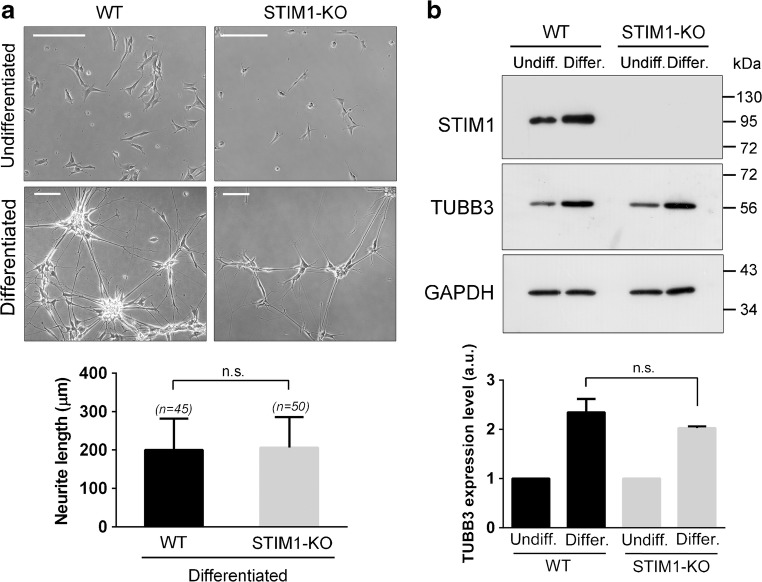
STIM1 deficiency did not modify markers of differentiation. a STIM1-KO cells and the parental cell line were differentiated as indicated above, and images of cells in culture were recorded to assess neurite length in undifferentiated cells (top panels), and differentiated after 12 DIV of treatment (bottom panels). Scale bar = 200 μm. Quantification of neurite length revealed no differences between wild-type and STIM1-KO cells. Data are presented as the mean ± s.d. of two independent experiments (n = 50 cells for KO; n = 45 cells for wt). b TUBB3 expression was studied by immunoblot, as in Fig. 2, using GAPDH as a loading control. Differentiation was stopped at 9 DIV and the relative expression of TUBB3 was assessed in three independent experiments (data are the mean ± s.d.)

