Fig. 5.
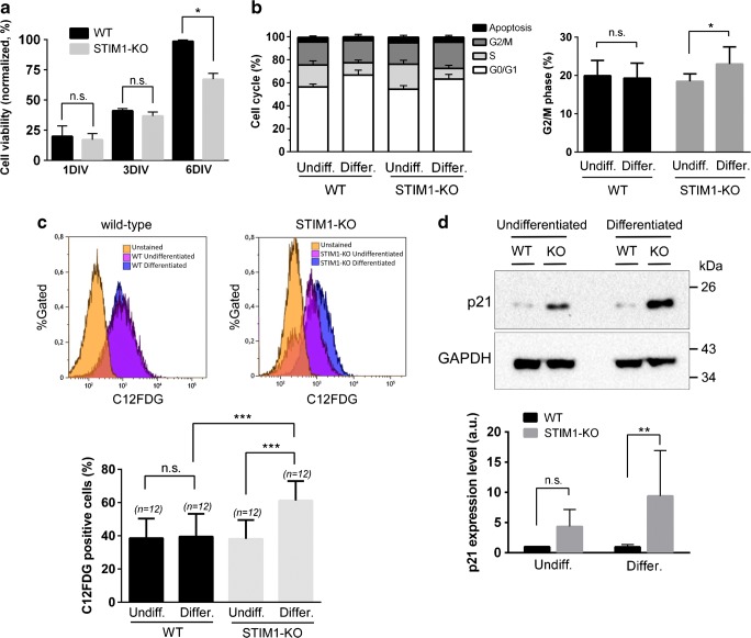
Loss of cell viability in differentiating STIM1-KO cells. a Cell viability of wild-type (black bars) and STIM1-KO cells (gray bars) was evaluated with an MTT assay at different stages of differentiation: undifferentiated cells, 24 h in growing medium (1 DIV); 24 h in growing medium + 2 days in differentiating medium (3 DIV); 24 h in growing medium + 5 days in differentiating medium (6 DIV). Data are presented as mean ± s.d. of three independent experiments, and results are normalized to the values obtained from wild-type cells at 6 DIV. b The analysis of the cell cycle was performed in undifferentiated (1 DIV) and differentiated cells (6 days of differentiation), and the assay was performed by staining fixed cells with propidium iodide and analyzing cells by flow cytometry. The percentage of cells at G2/M phase is plotted in the right panel to show the statistically significant increase of this phase in STIM1-KO differentiated cells. In both panels, data are the mean ± s.d. of three independent experiments. c Cells were cultured as indicated above, and stained with C12FDG to evaluate senescence by flow cytometry. The top panels show representative data histograms, with data from unstained cells as negative control (in orange), undifferentiated cells (pink), and differentiated cells after 6 DIV (blue). The y-axis is the normalized cell number, and the x-axis is the fluorescence intensity from C12FDG. Data of four independent experiments are shown in the bottom panel as mean ± s.d. d Lysates from cells in the experimental conditions described for panels (b, c) were assessed for p21 expression by immunoblot. The top panel shows a representative blot, with GAPDH as a loading control. The bottom panel shows data of four independent experiments as mean ± s.d.

