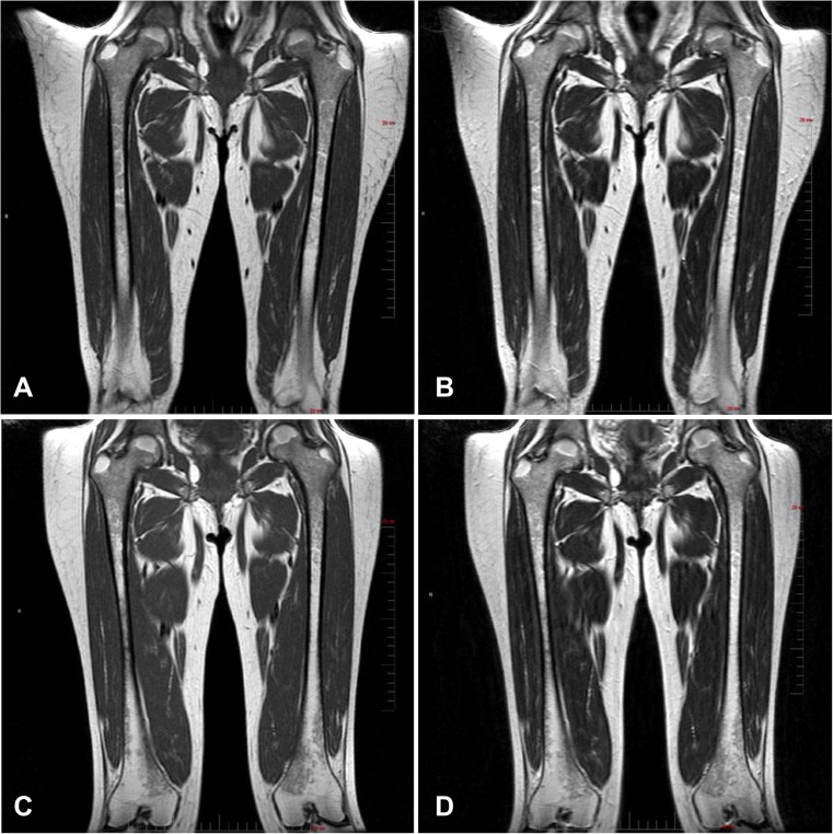
Fig. 3.
Assessment of olipudase alfa on bone marrow burden. Femur. Bone marrow burden changes in the coronal femur of patient 2, (female, 32 years old at baseline). Note the extent of hypointensity of the proximal epiphysis bone marrow at screening in the T1-weighted (a) and T2-weighted (b) images compared with the reduced amount and slightly hypointense diaphyseal bone marrow following 30 months of treatment (T1-weighted, c and T2-weighted, d). Full vertical scale bar, 20 cm. Spine. Bone marrow burden in the sagittal lumbar spine of patient 2. At screening, diffuse infiltration of the bone marrow is observed with T1-weighted isointensity of the non-diseased intervertebral discs (a) and T2-weighted hyperintense signal intensity of presacral fat (b). After 30 months of treatment, the infiltration of the bone marrow remains unchanged (T1-weighted, c) while the presacral fat is improved to slightly hyperintense (T2-weighted, d). Full vertical scale bar, 20 cm


