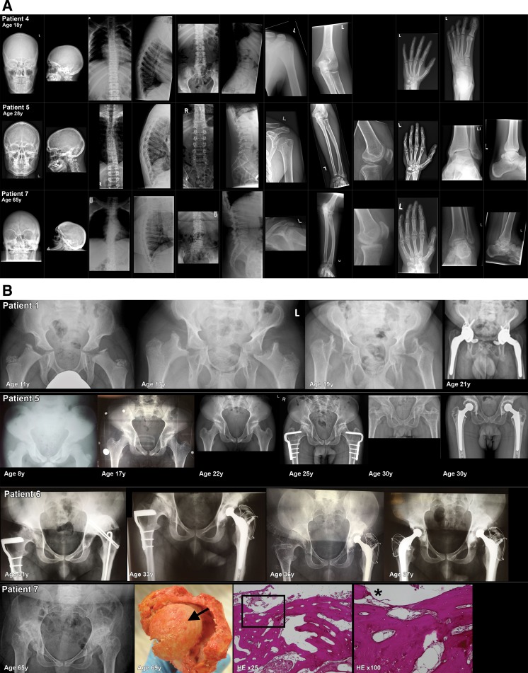
Fig. 1.
Skeletal radiographs. a Examples of three MLIII patients, aged 18, 28 and 65 years (patient numbers 4, 5, and 7). Skeletal radiographs of the skull (anterior posterior and lateral), spine (thoracic/lumbar vertebrae AP and lateral), left shoulder (AP), left elbow (lateral), left knee (AP), left hand (AP) and left ankle/foot (AP and or lateral). In general, the developmental bone abnormalities were present in all patients, but the presence and severity of osteoarthritic changes were more prominent in the older patients. Skull: In all patients, thickened cortical bones and a prominent sella turcica were observed. Open skull sutures in patient numbers 4 and 5. Dens aspect of the skull vault in patient number 7. Spine: Mild convex right-sided scoliosis, with increased kyphosis and increased interpedicular distances in all three patients. Flattened corpora vertebrae on several levels (cervical, thoracic and lumbar) in all three patients. Osteoarthritic changes of the endplates of the corpus vertebrae, most prominent in the oldest patient (patient number 7). In patient 7, there is anterior displacement of vertebrae L3 and L4 with a decreased diameter of the spinal cannel. Shoulder and elbows: In patient number 4, no abnormalities of these joints were observed. Osteoarthritic changes in the humeral head, glenoid and elbow deformation were seen in patient numbers 5 and 7. Knee: From patient number 4, no lateral left knee radiograph was available. In patient number 5, there is a patella baja and signs of osteochondral abnormalities of the patella with osteophyte formation. In patient number 7 (X-ray performed at age 60 years), osteoarthritic changes were observed with lateral hook formation/bone formation of the tibia plateau and at the lateral femur condyle. Hand: Abnormal shaped phalanges in all three patients (subtle in patient number 4). Osteoarthritic changes of the phalangeal joints (proximal and distal) in patient numbers 5 and 7. Abnormal shaped metacarpal bones (hypoplasia and collapse) with secondary osteoarthritis (patient numbers 5 and 7). Ankles/feet: In patient number 4, no abnormalities of the joints were observed. In patient number 5, there is osteoarthritis of the distal fibula. Suggestion for bifida talus or talus bipartite. Severe osteoarthritis of the ankle is seen in patient number 7. b Radiographs, macroscopy and histopathology of the hip bones. X-rays of the hips of patient numbers 1, 5, 6 and 7 over different ages. Macro- and microscopic photographs of the left hip of patient number 7 are shown. This patient died at the age of 69 years from metastatic bladder cancer. The most prominent findings on radiographs: Pelvis: In all patients, the pelvic bones are abnormally shaped, with flared iliac wings with hypoplasia of the inferior part of the ilea. The acetabula are severe dysplastic, very steep and shallow. Neoacetabulum formation occurred in patient numbers 5, 6 and 7. Impingement of the coxofemoral spaces was seen in patient number 7. Femoral heads, neck shaft angle: Severe ossification disorders and severe secondary osteoarthritis of the femoral heads (with subchondral cysts, sclerosis and flattening in patient numbers 5, 6 and 7) were present in all patients. In patient number 1, at age 11 years, there was near total absence of the femoral heads. Femoral shaft angle abnormalities; in patient number 1, the shaft angle over time develops from coxa valga to coxa vara. In patient number 5, there is a coxa valga and in patient number 7 coxa vara. On autopsy in patient number 7, part of the left femur, femoral head and part of the acetabulum were removed, shown on the macroscopic photo. The femoral head (shown from above); severe osteoarthritis is present, with complete destruction of the cartilage. An arrow on the top of the femoral head shows yellow coloured bone tissue and not the normal glossy blue-white in appearance cartilage. Total destruction of cartilage is also illustrated by the histological slides of the upper part of the femoral head coloured with HE, magnification ×25 and ×100. A square on the ×25 magnification indicates the location of the ×100 magnification. No cartilage remains at the location of the asterisk on the ×100 magnification. Surgical interventions of the hip: Patient number 1: custom-made total hip replacement (THR) of the left and right hips at 20 and 21 years, respectively. Patient number 5: femoral varus osteotomy at ages 22 years (left hip) and 24 years (right hip) and THR at age 30 years. Patient number 6: femoral varus osteotomy at ages 22 years (left hip) and 24 years (right hip) and THR at 31 years (left hip) and 37 years (right hip)

