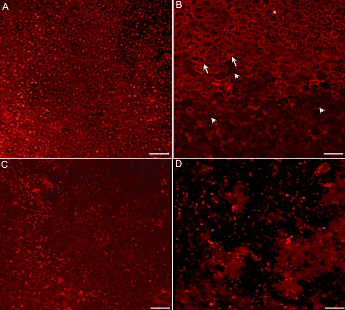
Figure 6.
Flatmounts of RPE stained with RPE65. (A) At 1 day after injection, RPE65 staining clearly outlines the cell membranes, showing the hexagonal shape of the RPE cells. Cells are intact and healthy. (B) At 3 days after injection, some atrophic and hypertrophic (arrows) and mottled (asterisk) RPE cells are observed as are RPE ghosts (arrowheads). (C) At 7 days after injection, the RPE cells are completely lost with only fragments of cells remaining in the injected area. (D) At 28 days, only RPE fragments remain in the injected area. Scale bars indicate 50 μm.

