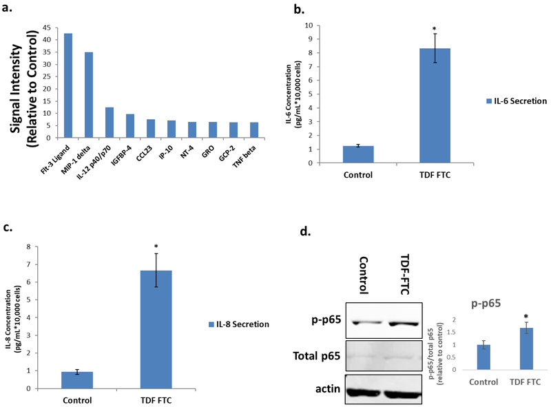
Figure 3. Secretory profile of HAART Drug Treated HUVECs.
HUVECs were treated with 10 μg/mL of tenofovir (TDF) and emtricitibine (FTC) for 7 days. (A) Senescence-associated secretory phenotype (SASP) analysis. After antiretroviral treatment, HUVECs were subjected to a 24-hour incubation in MCBD105 media to generate conditioned media (CM). The secretory profile was detected by incubating CM on a cytokine membrane array and normalized to cell number. Values are relative to a vehicle control. (B) IL-6 quantitation. CM was collected as in A and IL-6 was quantitated by ELISA. (C) IL-8 quantitation. CM was collected as in A and IL-8 was quantitated by ELISA. (D) Left, representative western blot illustrating protein levels of inflammatory mediator p65and loading control. Right, quantification. * = p value < .05, n = 3, error bars are S.D.

