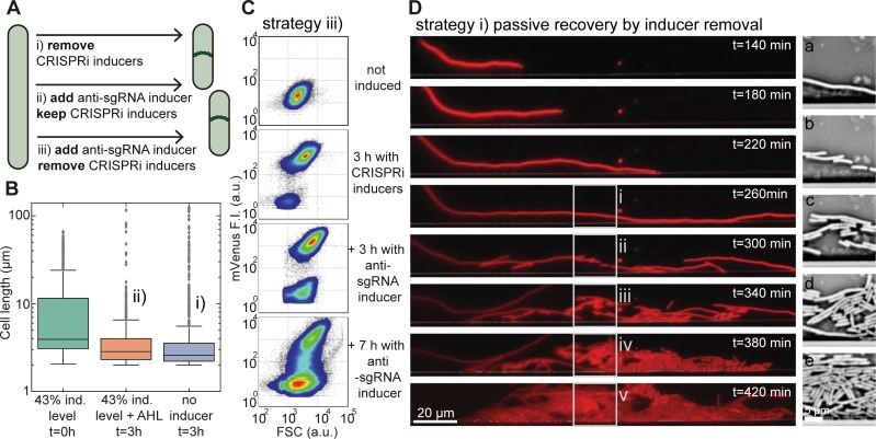
Fig 4. Restoration of cell division.
(A) Scheme showing three different strategies by which filamentous bacteria can be returned to normal cell division and growth. (B) Box plot of cell lengths (1000 cells each) of induced filamentous cells (at t = 0 hours) and their length after 3 h in medium supplemented with AHL (in the presence of IPTG, aTc), or after 3 hours in medium without any inducers. For both rescue strategies, the mean cell size shifted back from <L> = 10 μm to <L> = 4 μm. (C) Flow cytometer density plots of bacteria displaying mVenus fluorescence vs. forward scatter (FSC) signal from a non-induced, normal growth state (top) towards the filamentous state by induction with IPTG/aTc (second) and back again (bottom) by supplementation of AHL (in the presence of IPTG/aTc). Both the mVenus and FSC signals increase in the filamentous growth mode. After the release of anti-sgRNA, the size of the filamentous population decreases and recovers a normal cell morphology. (D) Left column: Fluorescence microscopy time series in the mRFP channel of an E. coli cell in a microfluidic chamber in the absence of inducers. Prior to the first image (top), the bacterium was grown filamentous for two hours at 43% induction level followed by a 140-minute exposure to growth medium without any inducers. At 300 minutes of growth, the bacterium resumes cell division. The new daughter cells quickly approach the normal cell size. Right column: images (a)-(e) show bright field images corresponding to the boxed regions on the left.

