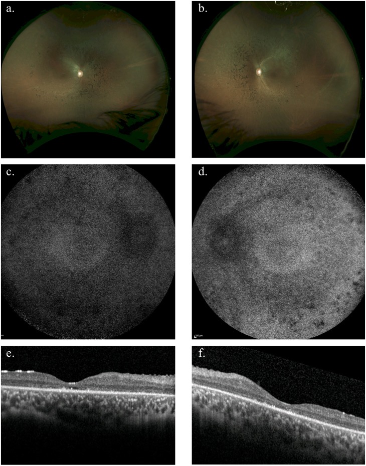
Fig 3. Fundus photographs and optical coherence tomography (OCT) for the affected individual in family 8.
Colour fundus photographs (a and b: OD and OS respectively) revealed attenuated retinal vessels, mid-peripheral coarse pigment clumping and white dots at the level of the retinal pigment epithelium. 55 degree fundus autofluorescence imaging (c and d: OD and OS respectively) show widespread loss of autofluorescence more marked over the pigment clumps in the mid-periphery. Optical coherence tomography (OCT) (e and f: OD and OS respectively) demonstrated loss of outer nuclear and photoreceptor layers throughout the macula with occasional small foci of retained photoreceptors. This individual presented with a history of night blindness from 1–2 years of age and an intermittent divergent squint with long-standing photophobia. Presenting visual acuity was 6/9 in each eye and at last review at age 18 years had deteriorated to 6/60 each eye with severely restricted visual fields to less than 15 degrees to confrontation. There was a myopic astigmatic refractive error of right 0.25/-1.25 x 21° and left 0.25/-2.25 x 160°. Electrophysiology performed at age 7 was unrecordable.

