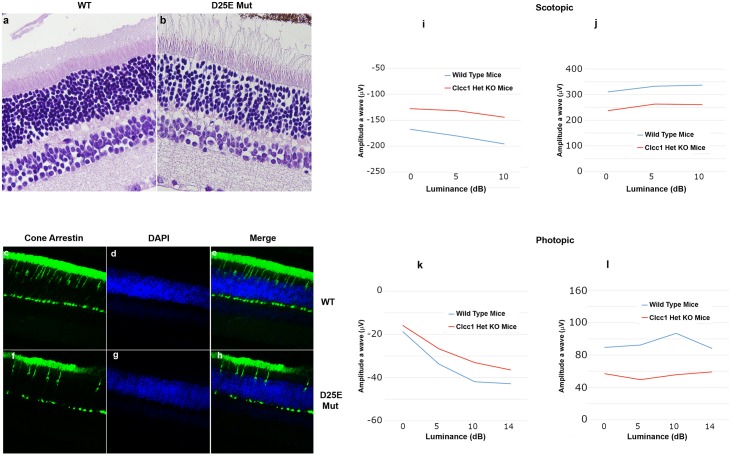
Fig 9. Effects of heterozygosity for Clcc1 KO on mouse retinas.
Hematoxylin & eosin staining of (a) WT and (b) Clcc1-/+ knockout 7-month-old mouse retinas. While the overall structure of the retina is preserved, staining revealed decreased cell density in the outer and inner nuclear layers, as well as the outer and inner plexiform layers as well as structural disarray of the photoreceptor layer in Clcc1 KO heterozygous compared with the WT mice. (c-h) Immunostaining of cone arrestin in WT (c-e) and heterozygous Clcc1 knockout (f-h) mice. The WT mice exhibit normal cone photoreceptors staining pattern while Clcc1 heterozygous KO mice revealed reduced number of cone photoreceptors. (i-l) Electroretinography of WT and Clcc1 knockout heterozygous mice show approximately 20–50% decreases in the amplitude of both the scotopic and photopic a and b wave amplitude responses in Clcc1 KO heterozygous mice compared to WT at all levels of luminance.

