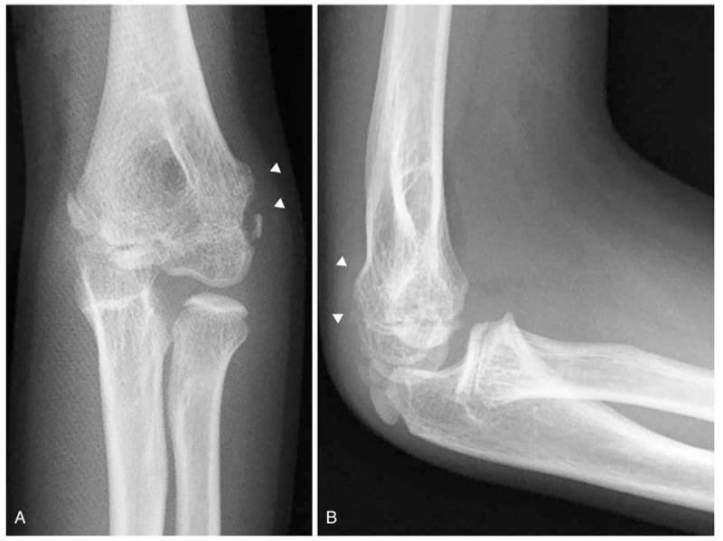
Figure 10.

Radiographs (A, B) of the left elbow in case 3, 16 months postoperatively showing decreased carrying angle in the elbow, and the anteroposterior view showing cubitus varus deformity. White arrowheads indicate the deformity of the distal humerus.
