Figure 8.

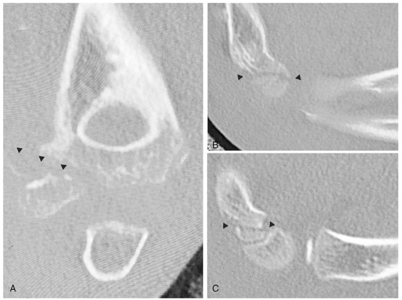
Computed tomographic images. Coronal image (A) and Sagittal images (B, C) of the left elbow in case 3 showing Milch type I lateral humeral condylar fracture. Black arrowheads indicate the fracture lines of the distal humerus.

Computed tomographic images. Coronal image (A) and Sagittal images (B, C) of the left elbow in case 3 showing Milch type I lateral humeral condylar fracture. Black arrowheads indicate the fracture lines of the distal humerus.