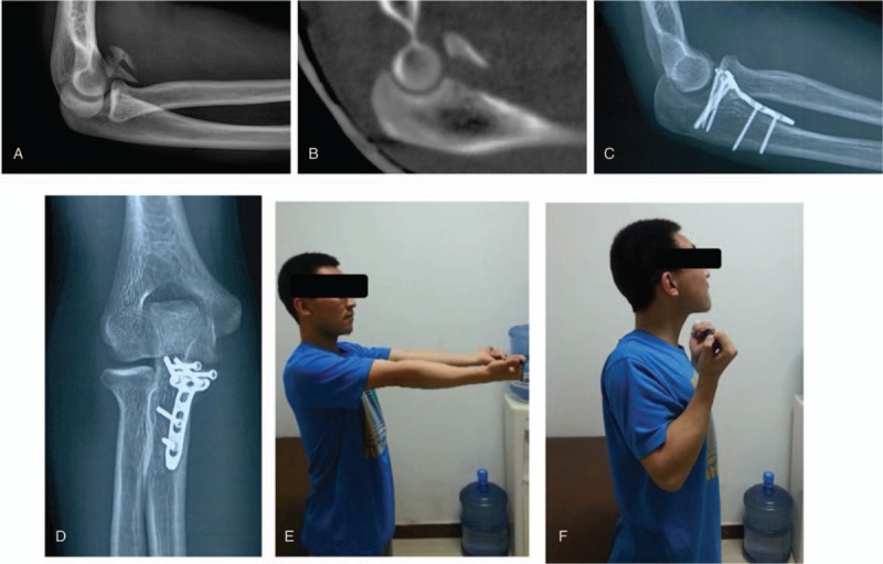
Figure 2.

Case example. A 17-year-old boy presented with a Regan and Morrey type III coronoid process fracture and fracture of the ipsilateral distal radius. Preoperative x-ray (A) and computed tomography (B) images show a severe comminuted type III coronoid fracture. Solid union and good outcome were achieved at the 8-month follow-up (C–F).
