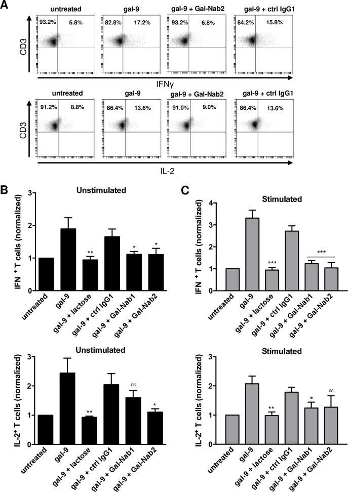
Fig 3. Anti-gal-9 mAbs inhibit the “Th1-like” phenotype shifting induced by gal-9 in resting or stimulated PBMCs.
A-C. PBMCs from healthy donors were stimulated or not with anti-CD3/CD28 antibodies and treated for one week with human recombinant gal-9 (Gal-9S; 40 nM) with or without combination with lactose (5 mM), control isotype antibody (ctrl IgG1), Gal-Nab1 or Gal-Nab2 (67 nM i.e. 10 μg/mL). Intracellular cytokine expression was assessed by flow cytometry as explained under “Materials and Methods”. At least 50% of the cells were alive. Dead cells were gated out. A. Examples of flow cytometry plots obtained with stimulated PBMCs for one donor after gating on the CD3+ T cell population: IFN-γ (upper plots) and IL-2 (lower plots) expression were analyzed. B-C. Percentages of CD3+ IFNγ+ (upper histogram) and CD3+ IL-2+ (lower histogram) cells were normalized with the basal percentages obtained in untreated cells using either unstimulated (B) or stimulated (C) PBMCs. Data are represented as means ± SEM of four independent experiments with different donors. All statistical differences displayed are compared with gal-9 treatment; ***p<0.001; **p<0.01; *p<0.05; ns: not significant (one-way ANOVA/Dunnet post-test).

