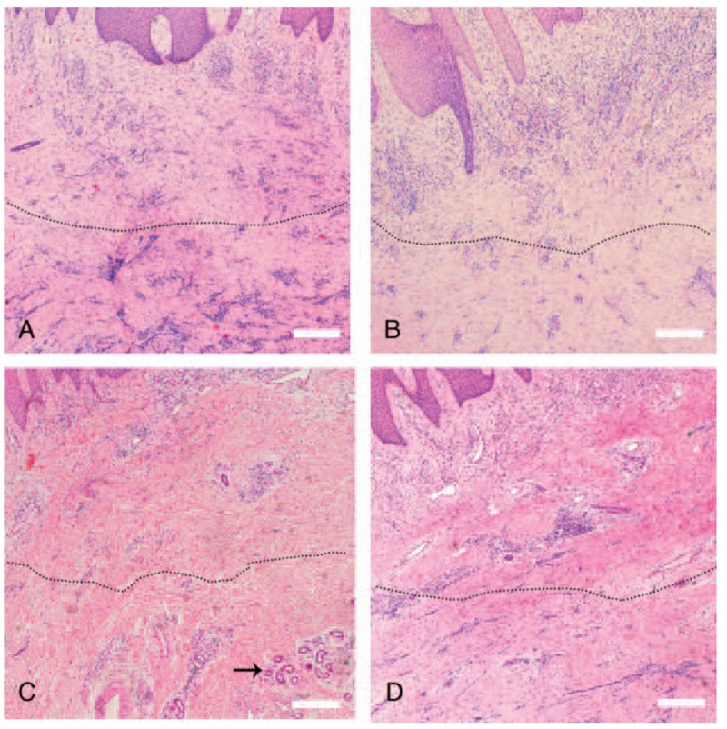
Figure 4.

Hematoxylin and eosin staining of tissue from the wound area. (A) Numerous inflammatory cells infiltrate the deep dermal layers (below dotted line) before treatment with ECM/SVF gel grafting. (B) Numerous inflammatory cells infiltrate the deep dermal layers (below dotted line) before negative pressure wound therapy. (C) Decreased inflammatory cell infiltration in the dermis deep layer (below dotted line) and new vascular structures (black arrow) after ECM/SVF gel grafting. (D) The inflammatory cell infiltration had not significantly changed (below dotted line) after negative pressure wound therapy. Scale bar=20 μm. ECM = extracellular matrix, SVF = stromal vascular fraction gel.
