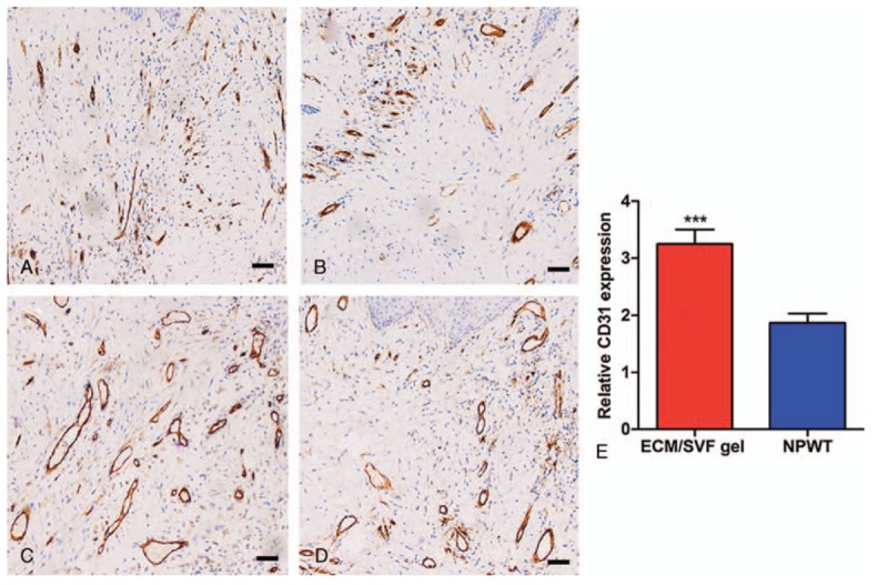
Figure 6.

Immunohistochemistry with CD31 of tissue from the wound area. (A) Scant neovascularization before ECM/SVF gel grafting. (B) Scant neovascularization before negative pressure wound therapy. (C) The greatest extent of neovascularization was seen after ECM/SVF gel grafting. (D) Neovascularization increased after negative pressure wound therapy. (E) Relative quantification of neovascularization. ∗∗∗P < .001. Scale bar=20 μm. ECM = extracellular matrix, SVF = stromal vascular fraction gel.
