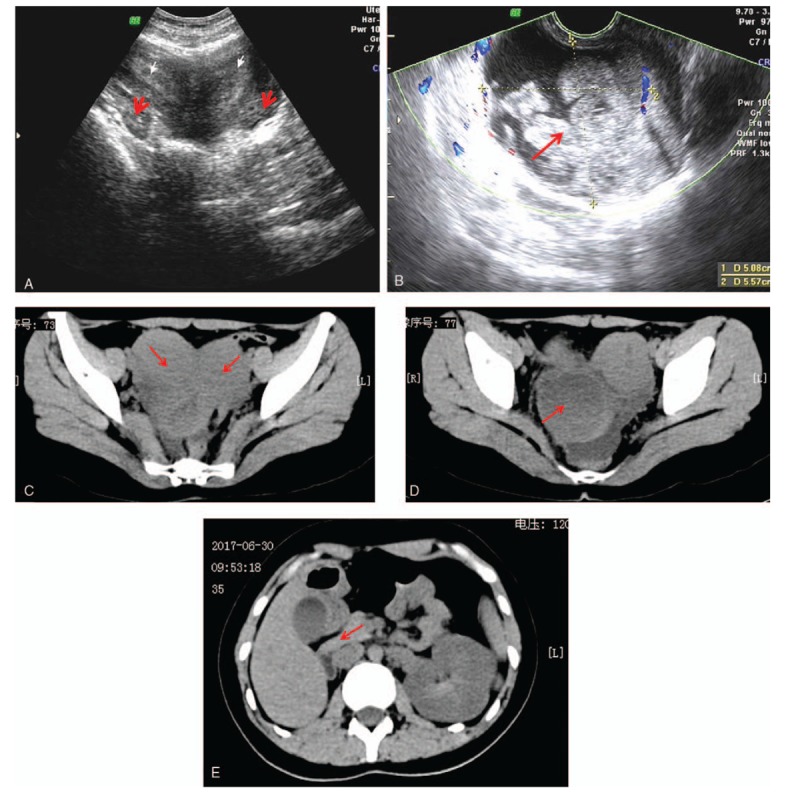
Figure 1.

(A) Transabdominal US showing uterus didephys (white arrow) and normal adnexal region (red arrow). (B) Transvaginal US showing hematocolpos mixed echogenicity (red arrow). (C) CT finding of uterus didelphys (red arrow). (D) CT finding of hematocolpos (red arrow). (E) CT finding of the right kidney is absent (red arrow). CT = computed tomography, US = ultrasound.
