. 2018 Aug 31;7:e40889. doi: 10.7554/eLife.40889

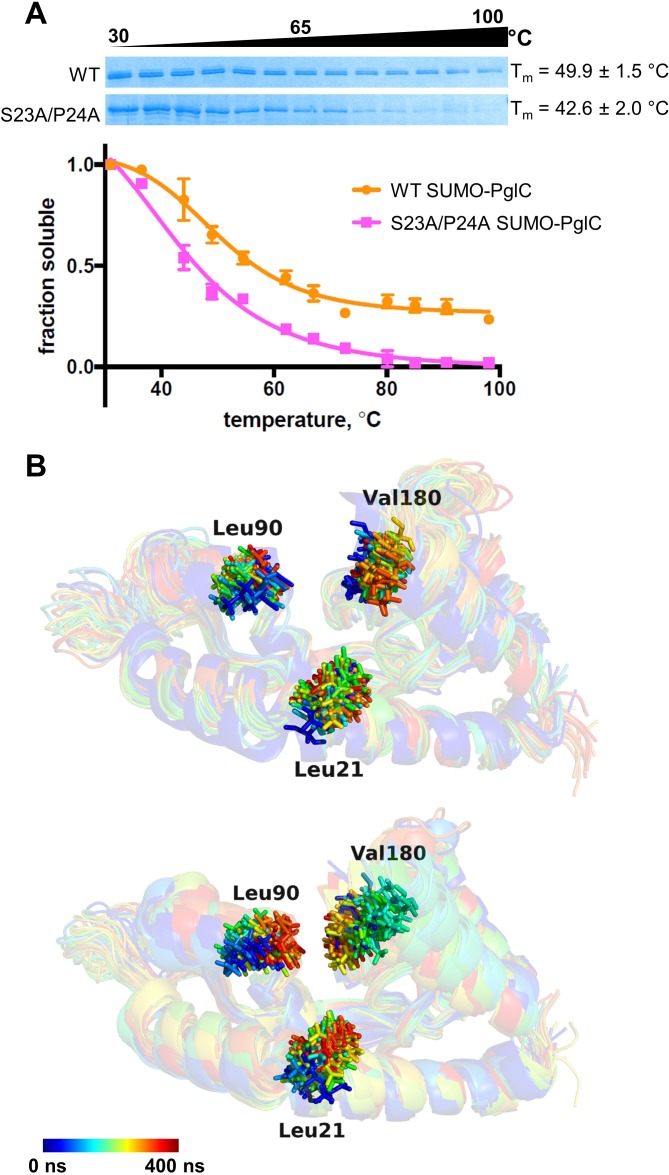
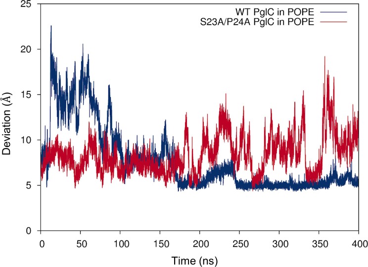
Figure 4. The Ser-Pro motif contributes to stability of the PglC fold.

(A) Thermal shift analysis of wild-type and S23A/P24A SUMO-PglC. Error bars are given for mean ± SEM, n = 3. (B) Superimposition of frames, taken at 10 ns intervals, along MD simulations of wild-type (top panel) and S23A/P24A (bottom panel) PglC. Colored from blue, t = 0 ns to red, t = 400 ns. PglC is represented as a semi-transparent cartoon and residues Leu21, Leu90 and Val180 as sticks.

Figure 4.

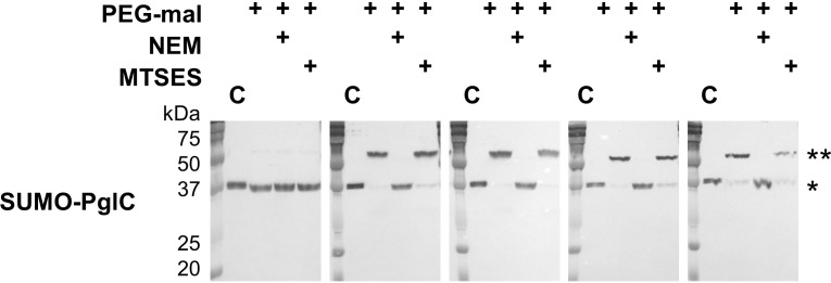
Figure 4—figure supplement 1. SUMO-PglC shows a reentrant topology similar to native PglC.

Figure 4—figure supplement 1.

SCAM analysis of wild-type SUMO-PglC topology.SCAM experiments were performed in duplicate or more. Representative Western blots are shown.
Figure 4—figure supplement 2. SDS-PAGE analysis of wild-type and S23A/P24A SUMO-PglC.

Figure 4—figure supplement 2.

Comparative SDS-PAGE analysis of wild-type and S23A/P24A SUMO-PglC, Coomassie stain; *=SUMO PglC. Sample loading was normalized by UV absorbance at 280 nm.
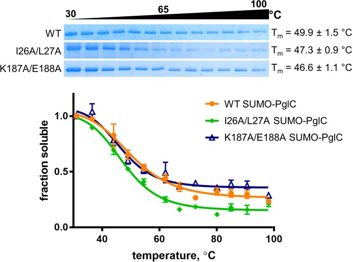
Figure 4—figure supplement 3. Thermal shift assays of control SUMO-PglC variants.

Figure 4—figure supplement 3.

Thermal shift analysis of wild-type, I26A/L27A and K187A/E188A SUMO-PglC. Error bars are given for mean ± SEM, n = 3.
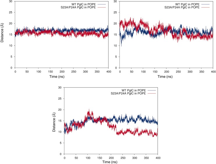
Figure 4—figure supplement 4. Mutation of the Ser-Pro motif causes a ‘collapse’ of the PglC fold interior.

Figure 4—figure supplement 4.

Cα-Cα distances between Leu21 and Leu90 (top, left panel), Leu21 and Val180 (top, right panel) and Leu90 and Val180 (bottom panel), measured over 400 ns of MD simulations of wild-type and S23A/P24A PglC in a POPE membrane.
Figure 4—figure supplement 5. Mutation of the Ser-Pro motif reduces lipid occupancy in the PglC fold interior.

Figure 4—figure supplement 5.

Distance between the geometric centers of the two lipids in the PglC fold interior, measured over 400 ns of MD simulations of wild-type and S23A/P24A PglC in a POPE membrane.