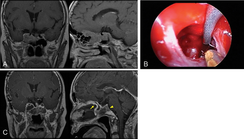
Fig. 1.

Recurrent nonfunctioning pituitary adenoma involving the cavernous sinus (CS). ( A ) Preoperative magnetic resonance imaging (MRI) demonstrating a pituitary adenoma in the right CS extending to the anterior cranial fossa. ( B ) Exposure of the internal carotid artery (ICA) in the right CS. The medial part of the CS tumor is being removed. ( C ) Postoperative MRI showing near total resection, with a small residual tumor (arrowhead) behind the right ICA. The arrow indicates a nasoseptal flap for the dural reconstruction.
