Fig. 7.

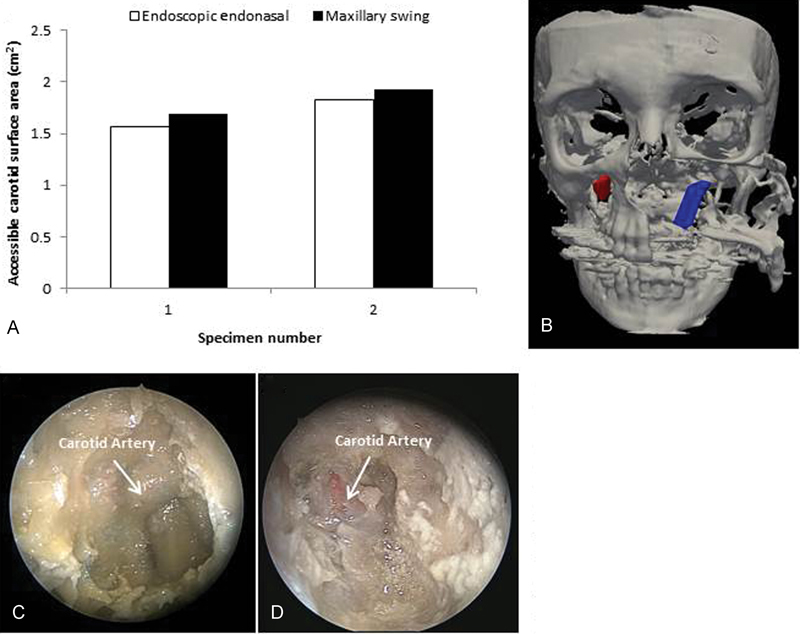
(A) Accessible internal carotid artery surface area using the endoscopic endonasal and maxillary swing approaches. (B) Surfaces of left and right internal carotid arteries traced by the optical tracking system. (C) The internal carotid artery as visualized during the endoscopic procedure. (D) The internal carotid artery as visualized during the maxillary swing procedure.
