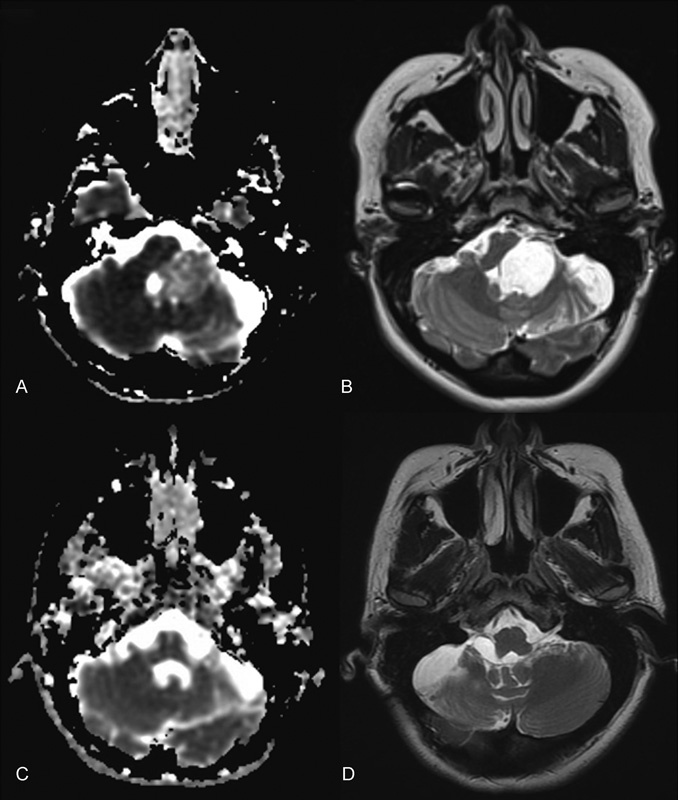
Fig. 1.

Axial diffusion-weighted image (A,C) and axial T2 magnetic resonance imaging (MRI) (B,D) showing large epidermoid cyst of the left cerebellopontine angle, brain stem, and foramen of Luschka before (A,B) and after (C,D) a gross total resection using a retrosigmoid approach. Intraoperatively, tumor was found compressing the fifth, seventh and eighth, and ninth and tenth cranial nerves. This patient presented to us after undergoing surgery at an outside hospital 9 years prior. Now 4 years after reoperation with routine MRI surveillance, there is no evidence of recurrence.
