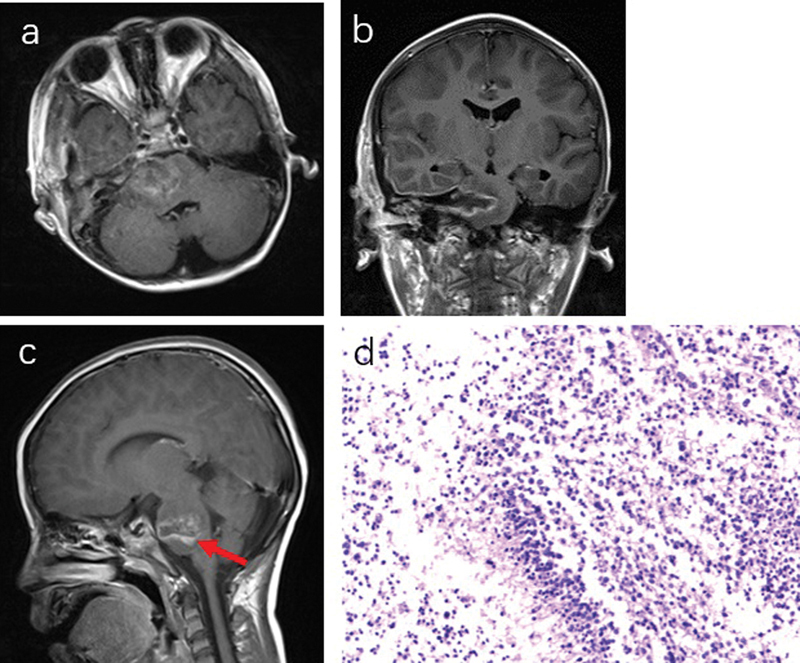
Fig. 4.

( a and b ) Postoperative MRI (3 days after operation) of patient 1. The tumor was extensive, but not completely removed ( c , red arrow), with the histological examination of glioblastoma ( d ) (WHO IV, GFAP + , Olig2 + , and K i -67, 60%). GFAP, glial fibrillary acidic protein; MRI, magnetic resonance imaging; WHO IV, World Health Organization grade IV.
