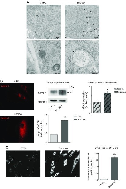
Figure 1.
Effect of sucrose loading on the endolysosomal compartment in human fibroblasts. A) Electron micrographs of human control (a, c) and sucrose-loaded fibroblasts (b, d). Arrowheads (b) and arrow (d) indicate sucrosomes; arrow (c) indicates lysosomes; N (a, b) indicates nucleus. B) Immunofluorescence staining of lysosomal marker Lamp-1 in permeabilized human fibroblasts, or not with sucrose (left). Immunostaining of Lamp-1 and loading control GAPDH accompanied with the semiquantitative graph of normalized Lamp-1/GAPDH (middle). Data are expressed as means ± sd. **P < 0.0021 vs. control (ctrl) (n = 4). Lamp-1 mRNA expression evaluated by quantitative RT-PCR (right). *P < 0.01 vs. ctrl (n = 3) Expression levels were normalized using HMBS and B2M as housekeeping genes. C) LysoTracker Red DND-99 staining performed on living human fibroblasts, loaded or not with sucrose, and a semiquantitative graph of the fluorescence intensity measured in ctrl and sucrose-loaded cells normalized by the number of cells analyzed. ***P < 0.0001 vs. ctrl (n = 70).

