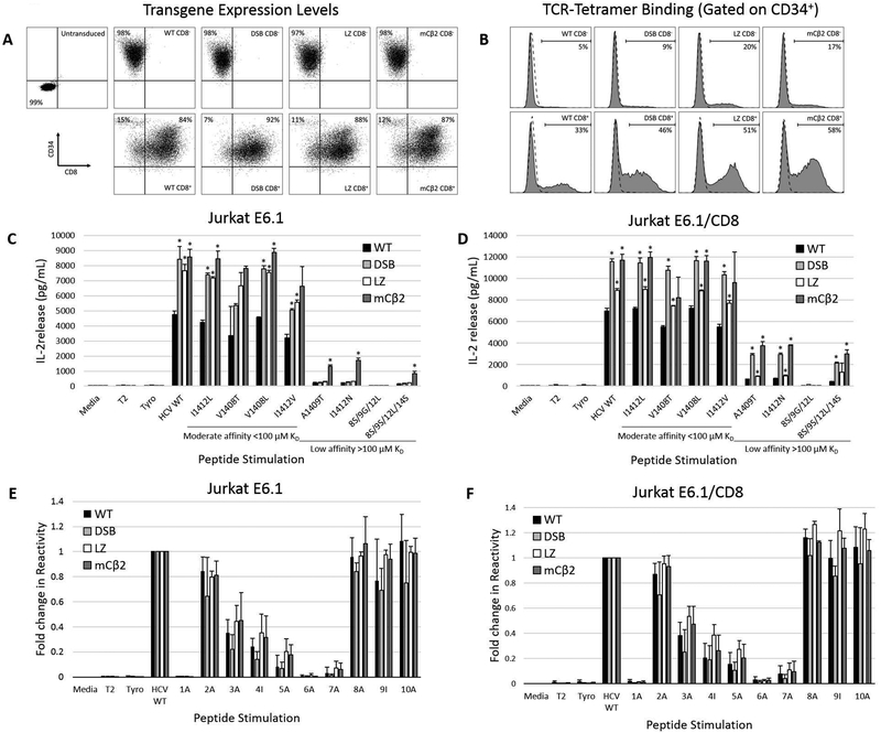
Figure 2. TCR pairing modifications enhance APL recognition by HCV1406 TCR-transduced Jurkat E6.1 cells.
(a) CD8- or CD8+ Jurkat E6.1 cells were engineered to express WT, DSB, LZ, or mCβ2 modified HCV1406 TCRs and immunomagnetically enriched for expression marker CD34 to ensure high and uniform transgene expression between groups. (b) Relative introduced HCV1406 TCR density was evaluated by tetramer binding. (c) CD8- or (d) CD8+ Jurkat E6.1 cells were co-cultured with WT and naturally occurring mutant NS3 peptide-loaded T2 cells. (e) CD8- or (f) CD8+ Jurkat E6.1 cells were co-cultured with WT and alanine-substituted NS3 peptide-loaded T2 cells. Fold change in cytokine release of each peptide substitution compared to WT sequence peptide is displayed. IL-2 release by WT (black bars), DSB (light gray bars), LZ (white bars), or mCβ2 (dark gray bars) TCR-transduced Jurkats was determined by ELISA. Mean and standard deviation of triplicate measurements are shown. APL are organized from by decreasing TCR-pMHC affinity, left to right. Significant differences in IL-2 release induced by modified TCRs compared to WT TCR are denoted *p<0.05. Data are representative of three independent experiments.

