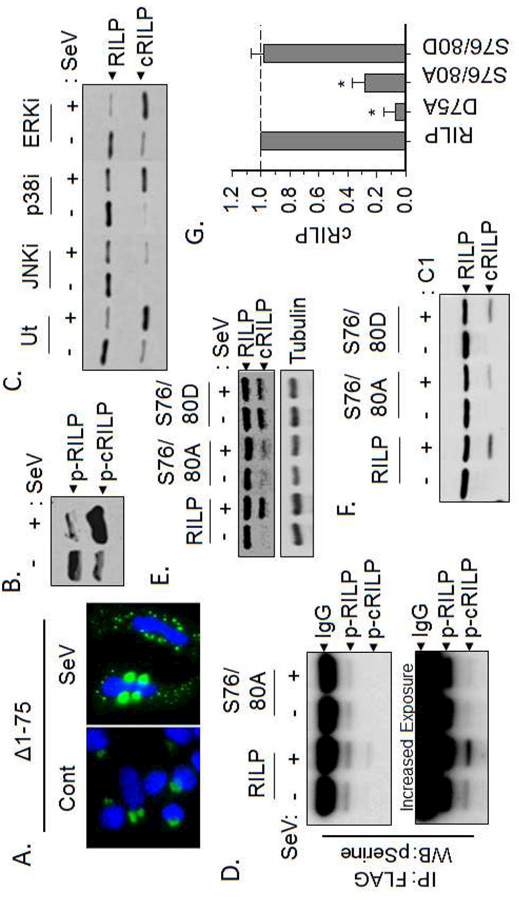
Figure 2. Efficient RILP cleavage requires phosphorylation.

(A) The cleavage mimic pEGFP-N1(RILP(∆1–75)) re-localizes only after Sendai virus infection. (B) Western blot analysis of total phospho-proteins showing that both total and cleaved RILP are phosphorylated however, the amount of phospho-RILP is greatly increased after SeV infection. (C) Inhibition of JNK, but neither p38 nor ERK, blocks RILP cleavage. (D) Western blot analysis of FLAG eluates for phospho-serine shows that cRILP is heavily phosphorylated after SeV infection. Mutation of the serines at positions 76 and 80 within RILP abolish this. (E) SeV induces the cleavage of wild-type RILP. The phospho-deficient RILP(S76/80A) blocks SeV-induced cleavage of RILP while the phosphomimetic RILP(S076/80D) efficiently and spontaneously cleaves. (F-G) Purified RILP protein was incubated with active caspase-1 as stated previously. Western blot analysis for RILP shows that phosphorylation-deficient RILP, S76/80A, is cleaved with less efficiency than wild-type or the phospho-mimic, S76/80D RILP.
