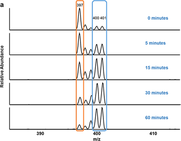
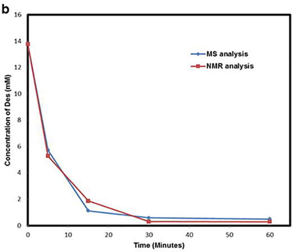
Fig. 5.


UV-induced degradation of Des- Degradation of Des was studied by irradiating the sample to UV light at 254 nm for different time intervals (0, 5, 15, 30 and 60 minutes). (a) Represents the MS2 spectra of UV-irradiated Des mixed with 25 ng/μL of Labeled-Des. The fragment ion generated from Des (decrease with time) and Labeled-Des are highlighted in orange and blue box respectively. (b) Represents the UV-induced breakdown by measuring concentration of Des at different time intervals using MALDI-MS2 and NMR spectroscopy
