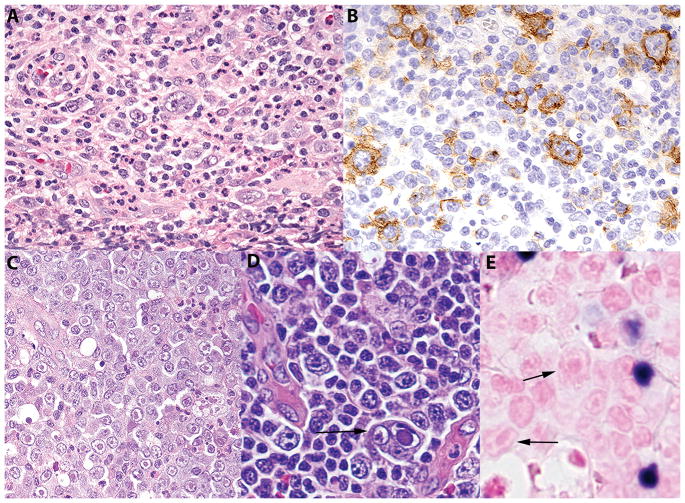
Figure 2.
Morphological and immunohistochemical features of CHL and AITL with HHV-6 infection. A, CHL, nodular sclerosis type with mononuclear Hodgkin cells and polymorphous background H&E ×400; B, CHL, the Hodgkin cells are highlighted by CD30 stain ×400; C, CHL, There were focal aggregates of cells with intranuclear eosinophilic viral inclusions similar in morphology to the viral lymphadenitis H&E, ×400; D, AITL, there were large atypical cells with intranuclear viral inclusions similar in morphology to the viral lymphadenitis. Arrow points a multinucleate cell with intranuclear inclusion H&E, ×400; E, AITL, there are small mononuclear EBV positive cells, which are clearly distinct from the virally infected cells (arrows), EBER-ISH, ×400. CHL – classic Hodgkin lymphoma, AITL – Angioimmunoblastic T cell lymphoma, EBER-ISH – Epstein Barr Virus early antigen – in situ hybridization.

