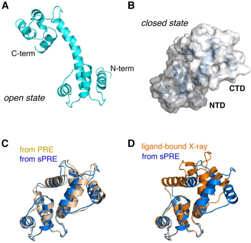
Fig. 7. A two-conformer ensemble structure identified for ligand-free Ca2+-CaM.

A) The open state selected from MD library with a population of 60%. B) The close state selected from MD library with a population of 40%. The two domains, with the NTD and CTD colored in different shades of gray, bury solvent accessible surface area of ~1255 Å2, while at the same time, the linker residues improve their solvent exposure at one side of the protein. C) Comparison of the closed-state structure identified based on the sPRE data (colored blue) and previously determined based on the PRE data with a nitroxide probe covalently attached at S17C site (colored light orange). The backbone r.m.s. difference between the two structures is as low as 1.50 Å. D) Comparison of the closed-state structure based on the sPRE (marine) and the crystal structure of the ligand-bound Ca2+-CaM (PDB code 1CDL, colored orange). The backbone r.m.s. difference between the two structures is as low as 3.50 Å.
