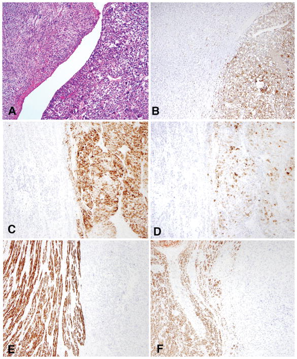
Figure 5.
In case 11, spindled cells with striking smooth muscle-like differentiation (left) co-existed with epithelioid cells (right) (A). The epithelioid component (right) was strongly positive for cathepsin K (B), HMB-45 (C), and melan-A (D), but negative for desmin (E) and h-caldesmon (F). The inverse staining pattern was noted in the spindled component.

