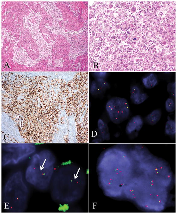
Figure 6.
In case 14, epithelioid clear cells with alveolar and nested patterns (A) were strongly positive for TFE3 by immunohistochemistry (B) with PSF and TFE3 rearrangements by FISH (C), as is typically seen in TFE3-rearranged PEComas. In case 12, diffuse sheets of highly atypical and mitotically active epithelioid cells (D) showed unbalanced complex RAD51B (E) and OPHN1 (F) gene rearrangements by FISH.

