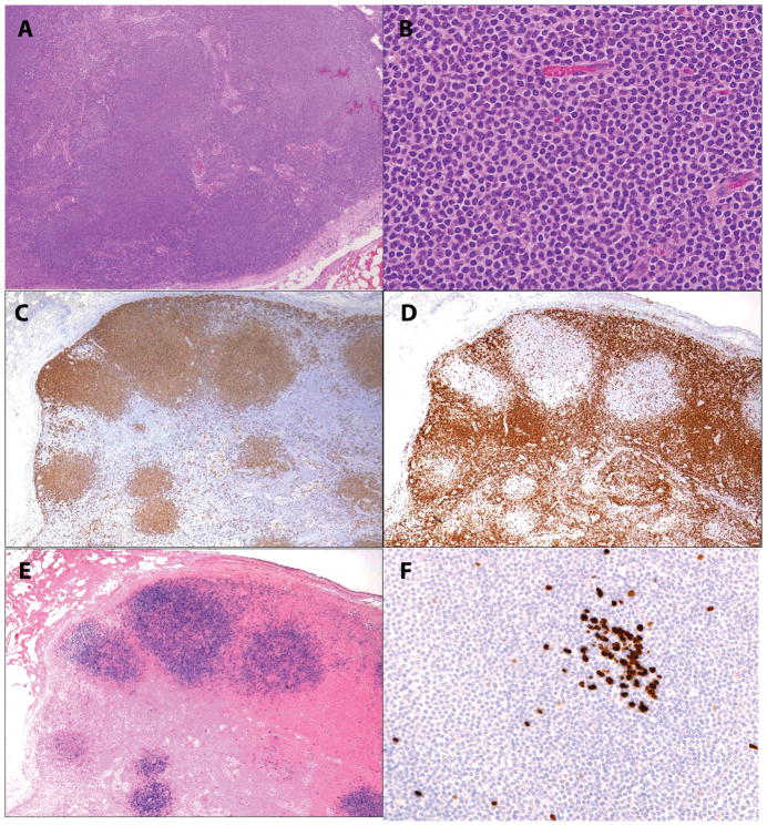
Figure 1.
EBV-positive nodal marginal zone lymphoma in an 18-year old female with heart/kidney combined transplant (case 1). A and B, Nodal architecture is effaced by nodular infiltrates of monocytoid cells with a rim of pale cytoplasm C. Monocytoid cells are positive for CD20, while CD3 [D] surrounds and delineates the tumor nodules. E. EBER shows a similar distribution to CD20. F. Ki-67 demonstrates a very low proliferation index in the monocytoid cells, while being positive in a small residual germinal center.

