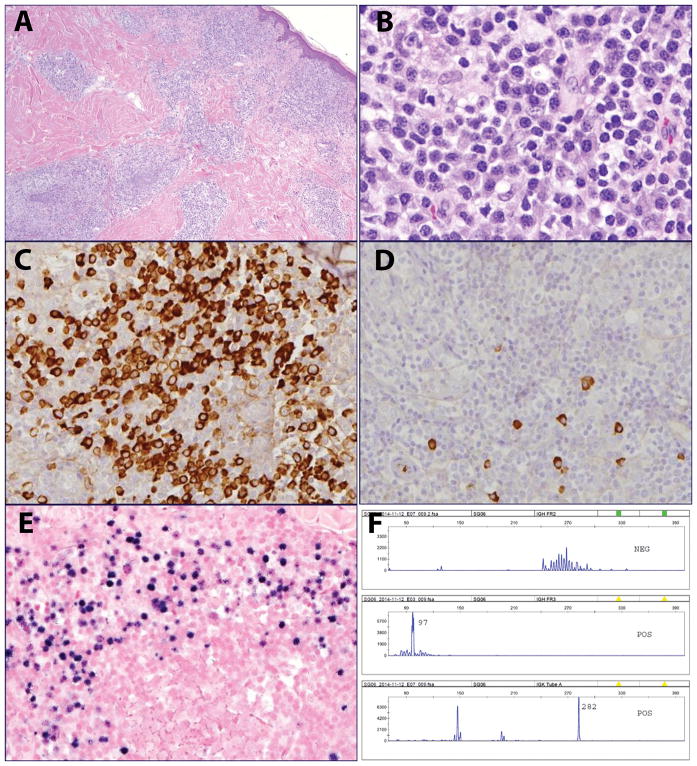
Figure 3.
EBV-positive MALT lymphoma in a 70-year old male with no significant past medical history (case 3). A. The infiltrate involves the dermis and subcutaneous tissue with a perivascular and periadnexal distribution. B. The infiltrate is composed of monocytoid and plasmacytoid cells. C. The plasmacytoid cells are positive for lambda and D. negative for kappa light chain. E. EBER shows a similar distribution to lambda. F. PCR for immunoglobulin gene rearrangements revealed a clonal pattern, with peaks in IGH Fr III, and Kde.

