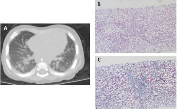
Figure 2. Liver biopsy and high resolution chest computed tomography.

(A) Diffuse infiltrative opacification in the lung periphery indicating severe interstitial lung disease. (B) H&E stain of liver section showing portal and sinusoidal fibrosis, cholangiolar proliferation and diffuse macrovesicular steatosis with ballooning of hepatocytes. (C) Masson trichome stain demonstrating portal and sinusoidal fibrosis.
