
Figure 1.
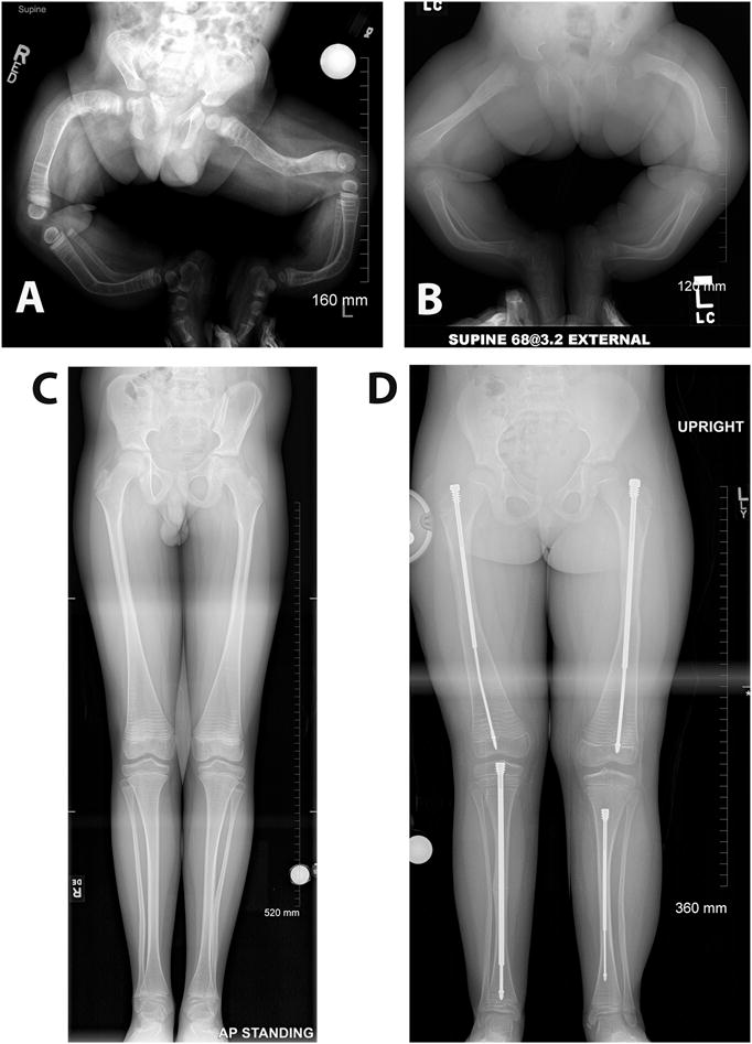
Radiographic images of lower extremities of patients affected with different types of OI, demonstrating the wide range of severity that affects the skeleton in this disease. A. One-year-old infant diagnosed with severe type III OI. Note the severe bowing of the legs and the lack of bone modeling in both femurs and tibiae. B. A nine-month-old infant with moderately severe type IV OI. C. A 12-year-old patient with type I OI. Note the relative constriction of the femur diaphysis and the flaring of the distal metaphysis which is known as Erlenmeyer flask deformity. Metaphyseal bands due to cyclical bisphosphonate treatment are also visible. D. Same patient shown in B at 7 years of age and after years of medical and surgical management. Note the telescoping rods implanted in both femurs and tibiae, a common surgical procedure in OI patients that have significant long bone deformities. All the radiographic images are a courtesy of Dr. Paul W. Esposito and Maegen Wallace (University of Nebraska Medical Center).