
Figure 2.
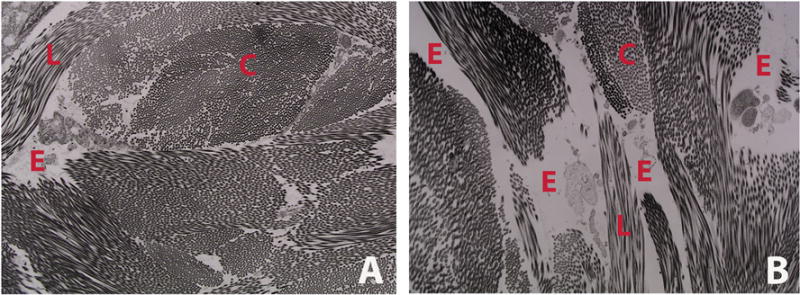
Representative transmission electron micrograph of dermal collagen from WT (A) and CrtapKO (B) mouse, showing decreased amount of collagen and increased space among collagen bundles in this murine model of recessive OI. This translates into skin laxity and reduced dermal thickness. C = cross section of collagen fibrils; L = longitudinal section of collagen fibrils; E = empty space.