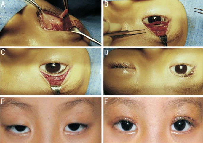
Figure 1. Procedure of frontalis muscle flap suspension surgery.

A: A 15×20 mm quadrangular frontalis muscle flap was dissected and pulled down; B: The free flap border was then attached to the tarsal plate with three 5-0 silk sutures and the redundant muscle was trimmed; C: Reset of the orbicularis oculi muscle; D: The eyelid level was adjusted according to the marking before anesthesia and the skin was closed; E, F: An example figure of a patient before and 1wk after the surgery.
