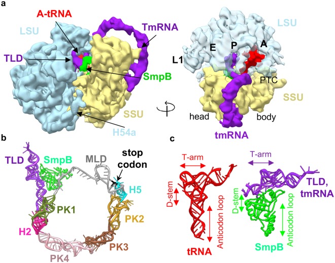
Figure 3.
Structure of the trans-translating state of Ms 70S ribosome. (a) Cryo-EM map of the tmRNA-SmpB complex bound 70S ribosome at 12.5 Å resolution. Density is shown as surface. tmRNA is shown in purple, SmpB in green, and A-tRNA in red (left). LSU in transparent blue showing the tmRNA-TLD and SmpB occupying the P-site and tRNA occupying A-site (right). (b) Architecture of tmRNA and SmpB when bound to the 70S. TLD is shown in purple, psuedoknots (PK) 1–4 are in deep green, pink, brown, and yellow, respectively. Helix 2 (H2) is in magenta, helix 5 (H5) in light blue, MLD in grey (stop codon in black), and SmpB in green. (c) Comparison of the structure of a typical tRNA with tmRNA-TLD and SmpB complex showing how tmRNA (TLD) and SmpB complex mimics a tRNA.

