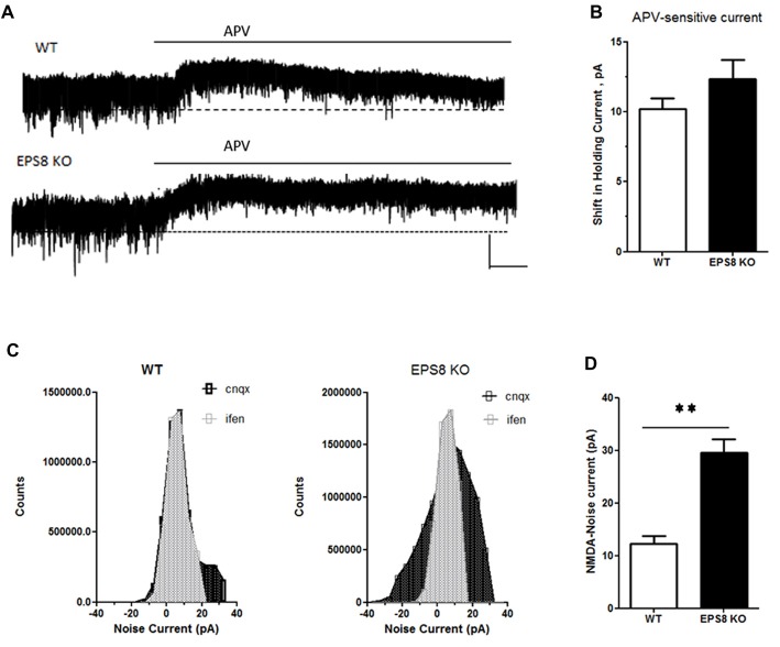
Figure 4.
Eps8 KO neurons display an increased NMDA-mediated tonic inward noisy current. (A) Examples of NMDA-only mEPSCs/“noise” recorded in [0 Mg2+]e ACSF which is abolished by perfusion with the NMDAR antagonist, APV. Dashed lines indicate extrapolated pre-APV shift in holding current. (B) Summary of the shift of holding current as a result of 50 μM APV application. (C,D) AP-5-associated change in a baseline noise. Quantification of background noise was obtained plotting all values of recordings traces and comparing the fluctuation of values distribution. Data are mean ± SEM (Mann Whitney test, **P < 0.01).

