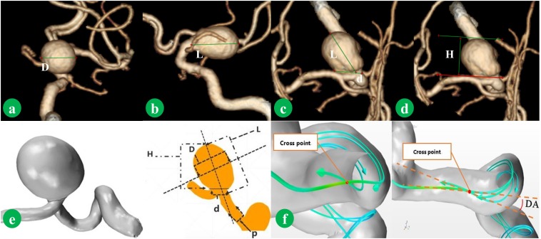
FIGURE 1.
Morphological parameters were measured from CTA image. D is the maximum diameter of the body (a). L is the maximum distance of the dome from the neck plane (b). d is the average diameter of the neck (c). H is the maximum perpendicular distance of the dome from the neck plane (d). The method of measurement is shown in (e). p is average diameter of the parent artery. Normal vector was combined. The deviated angle (DA), which was between co-velocity and normal vector, was measured (f).

